The following article about shivers in horses is reprinted from our sister publication EquiManagement. That is a brand created for equine veterinarians and experienced horse owners. The article reports on a collaborative 2023 study.1 The participants were equine practitioners from Michigan State University, Colorado State University, and the University of California, Davis. They examined histopathology samples of brain tissue from affected horses to try to identify the condition’s source.
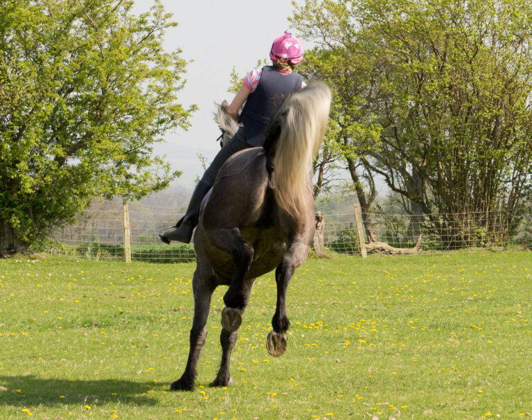
Horses with shivers often have difficulty walking backward or lifting a hind limb. This is because of fixed, hyperflexed abducted hind limbs or rigid hind-limb extension. This neuromuscular disorder is also accompanied by trembling, or “shivering,” of the tail and thigh muscles—hence, the moniker “shivers.” Affected horses tend to be able to move forward at walk, trot, and canter without problem until the syndrome progresses to hind-limb hyperflexion at the walk. Notable signs begin by 7 years of age.
Study on Source of Shivers in Horses
Previous study results identified selective degeneration of Purkinje cell (PC) axon projections in the lateral deep cerebellar nuclei. This is significant because the cerebellum is critical for “coordination of locomotion through integration of sensory information from the spinal cord and feedback from the motor cortex, basal ganglia, and thalamus.” Transmission of this information enables the PC to send efferent inhibitory outputs to motor pathways. “Walking backward, compared with walking forward, is more specifically influenced by cerebellar activities because the cerebellum exerts anticipatory postural adjustments in the absence of the ability to visually monitor steps,” the authors noted.
This study investigated a cerebellar cause of shivers. Other signs of cerebellar dysfunction are not considered clinical signs of shivers. These include proprioceptive deficits, truncal sway, and intention tremor, .
Study Population
In the current study, the research team hypothesized that horses with shivers have a distinct pattern of gene expression within the cerebellar white matter. They compared differences in gene expression between affected and control horses.
The study involved horses taller than 16.3 hands. The horses included three Belgian drafts, one Thoroughbred, and one Appendix Quarter Horse. The horses were age 7-13. All experienced shivers signs, such as hyperflexion of both hind limbs when walking backward and reluctance to lift a hind limb. They had had normal gaits at walk and trot. Other neurologic examination findings were normal.
The four control horses were taller than 16.2 hands and included three Thoroughbreds and one Irish Sport horse. The control horses had other issues, such as persistent lameness, a sinus tumor, or chronic back pain, but no evidence of neurologic disease. The control horses did not resist lifting a hind limb and moved normally both forward and backward.
All nine horses had been donated and were euthanized humanely to obtain, process, and analyze brain tissue.
Research Findings
The authors reported that “within the lateral cerebellar hemispheres, both spatial gene and protein expression differ significantly between shivers and control horses.” The lateral cerebellar hemispheres are critical to “motor planning and timing and onset and coordination of movements.” Differentially expressed genes (DEG) occurred in high numbers in cerebellar axons of shivers horses compared to control horses.
The researchers found that PC soma resists injury. PC axonal damage is not associated with ataxia or tremors, and shivers horses do not typically display these clinical signs. A neuroinflammatory cascade results in neurodegeneration and motor dysfunction in shivers horses by suppressing inhibitory GABA (gamma aminobutyric acid) receptor activities to decrease the number of Purkinje cells within cerebellar white matter.
Axonal injury also causes reduced expression of neurofilaments that affect the growth and stability of myelinated axons and enable optimal conduction velocity of electrical impulses along axons.
The study authors identified “decreased expression of proteins involved in myelinating axons in shivers horses.” They concluded that, “Our finding of altered expression of neurofilaments, myelin, and ion pumps and channels in the cerebellum of horses with shivers implicates an abnormality in axons in horses with shivers that could impact the cerebellum’s ability to coordinate movement.”
Further, shivers horses’ brain tissue has increased expression of calcium-dependent phospholipid binding protein (annexin). That is associated with inflammation and neurodegeneration. A number of other proteins related to heat shock, oxidative stress, calcium buffering or signaling, and actin had increased expression in the shivers horses in this study. Reduced expression of other proteins is associated with impaired motor coordination. Some of these are sex-specific. This is consistent with the fact that male horses are three times more likely to develop shivers than females.
In conclusion, the authors reported that Purkinje cell axonopathy is a cause of shivers in horses.
Reference
- Cerebellar axonopathy in Shivers horses identified by spatial transcriptomic and proteomic analyses. Valberg, S.J.; Willams, Z.J.; Henry, M.L.; Finno, C.J. Journal of Veterinary Internal Medicine. May 2023; DOI: 10.1111/jvim.16784
Further Reading
- Diseases and Conditions Archive. MySeniorHorse.com
- Additional Behavior and Musculoskeletal Pain Signs in Ridden Horses. Dr. Sue Dyson. MySeniorHorse.com
-
Nancy S. Loving, DVM, was a performance horse horse veterinarian based in Colorado for most of her career. She has a special interest in helping horse owners understand technical veterinary topics and research.View all posts


