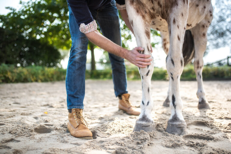
It is important to interpret radiographs appropriately to determine the likely clinical significance of radiological abnormalities of horses’ neck vertebrae.
An ‘abnormality’ on an X-ray (a radiological abnormality) is not necessarily synonymous with pain or dysfunction either currently or in the future. For example, radiological abnormalities consistent with osteoarthritis do not necessarily cause pain.
Neck Radiographs
Recent developments in radiographic equipment have made it possible to acquire neck X-rays in the field. However, interpretation of the radiographs is not straightforward for several reasons. Here are things that the veterinarian can do.
- Appropriately expose radiographs.
- There should have been no movement during image acquisition.
- Appropriately align the neck and X-ray beam. Misalignment can cause obliquity of the images, which prohibits accurate interpretation.
Control of these factors is easier said than done.
Clinical Significance
It is also important to recognise what abnormalities are of unlikely clinical significance. Loading and stressing bones can alter their shape and size. Given the large range of motion at the base of a horse’s neck (think about a horse raising its head on the approach to a jump or on landing versus slowly lowering the head and neck to graze), it is not surprising that modelling changes (alterations in size and shape) of the articular processes are common.
Moreover, such changes are not necessarily indicative of osteoarthritis.
Recent Study
A recent large-scale study evaluated radiographs of the fifth cervical to second thoracic vertebrae in Warmblood horses with neck-related clinical signs (neck pain and/or stiffness, neck-related forelimb lameness, or incoordination and weakness) and control horses with no such neck-related problems.
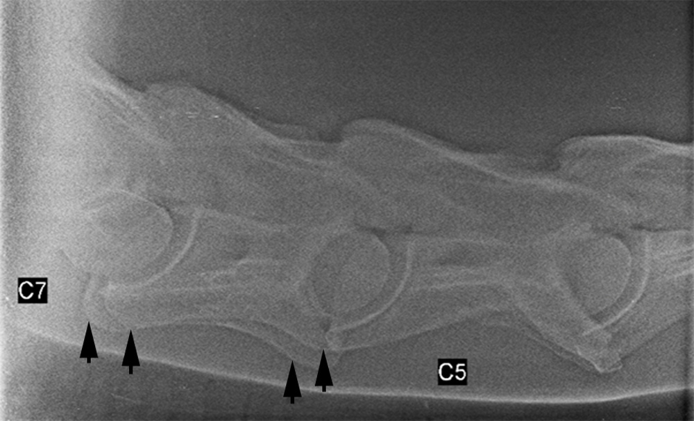
Severe modelling of the articular processes between the sixth and seventh cervical vertebrae or the seventh cervical and first thoracic vertebrae was more likely in cases compared with controls.
Mild and moderate modelling, however, were common in both control horses and cases. Interpret with care those cases.
Cases were more likely to have subluxation (malalignment) of the sixth and seventh cervical vertebrae and/or the seventh cervical and first thoracic vertebrae than control horses.
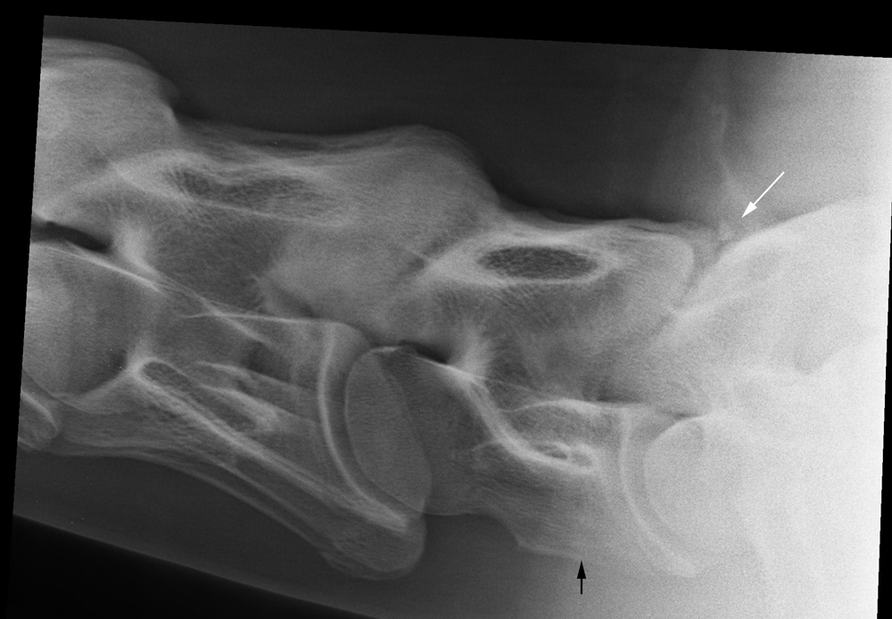
Abnormalities involving the region of the intervertebral discs with or without the adjacent vertebral bodies were more common in cases than control horses. Those involved articulations from the sixth cervical to second thoracic vertebrae.
The results highlighted the range of radiological abnormalities that can be seen in horses without associated clinical signs. This reinforces the necessity of careful clinical appraisal. The presence of radiological abnormalities does not imply a neck-related problem.
Final Words
Horses with neck-related clinical signs may have radiological abnormalities that are restricted to—or include—the first and second thoracic vertebrae. Significant lesions may be missed if the radiographic examination of horses with neck-related clinical signs is limited to the cervical vertebrae. It should include the first two thoracic vertebrae, .
Complete radiological assessment of the cervicothoracic region is important for accurate diagnosis and prognosis and for the development of treatment and management strategies.
Resource
Radiological abnormalities of the cervicothoracic vertebrae in Warmblood horses with primary neck-related clinical signs compared with control horses. Dyson, S., Quiney, L., Phillips, K., Zheng, S., Aleman, M. Vet Radiol Ultrasound 2024, 65: 755-768. doi: 10.1111/vru.13420
Further Reading
- Are Horses Born with Neck Vertebrae Variants Prone to Health Issues? Dr. Sue Dyson. MySeniorHorse.com
- Clinical Features of Primary Neck Problems in Horses. Dr. Sue Dyson. MySeniorHorse.com
- My Senior Horse Podcast: Horses and the Science of Harmony. Dr. Sue Dyson. MySeniorHorse.com
- Ridden Horse Performance Checklist: Behaviors in Ridden Horses that Might Signify Discomfort. Dr. Sue Dyson. MySeniorHorse.com
- Horse Behavior During Tacking and Mounting. Dr. Sue Dyson. MySeniorHorse.com
- How an Uncomfortable Horse Might Feel to a Rider. Dr. Sue Dyson. MySeniorHorse.com