Radiographs (X-rays) are commonly used in senior horses to help diagnose and monitor a range of conditions, from aching joints to dental disease. Like all imaging techniques, they have advantages and limitations. It is useful for senior horse owners to understand what an X-ray can and cannot show and what to expect if their horse needs X-rays.
What Is Radiography?
Radiography (or taking X-rays) is a painless, noninvasive imaging technique that uses electromagnetic radiation to create pictures of the internal and external structures of your horse’s body. The amount of radiation used in each X-ray is very small, and X-rays have been used safely for many decades in human and veterinary medicine. Veterinarians and nurses wear protective clothing whilst taking radiographs because they often X-ray many patients a year, potentially exposing themselves to much higher levels of radiation than with each patient alone.
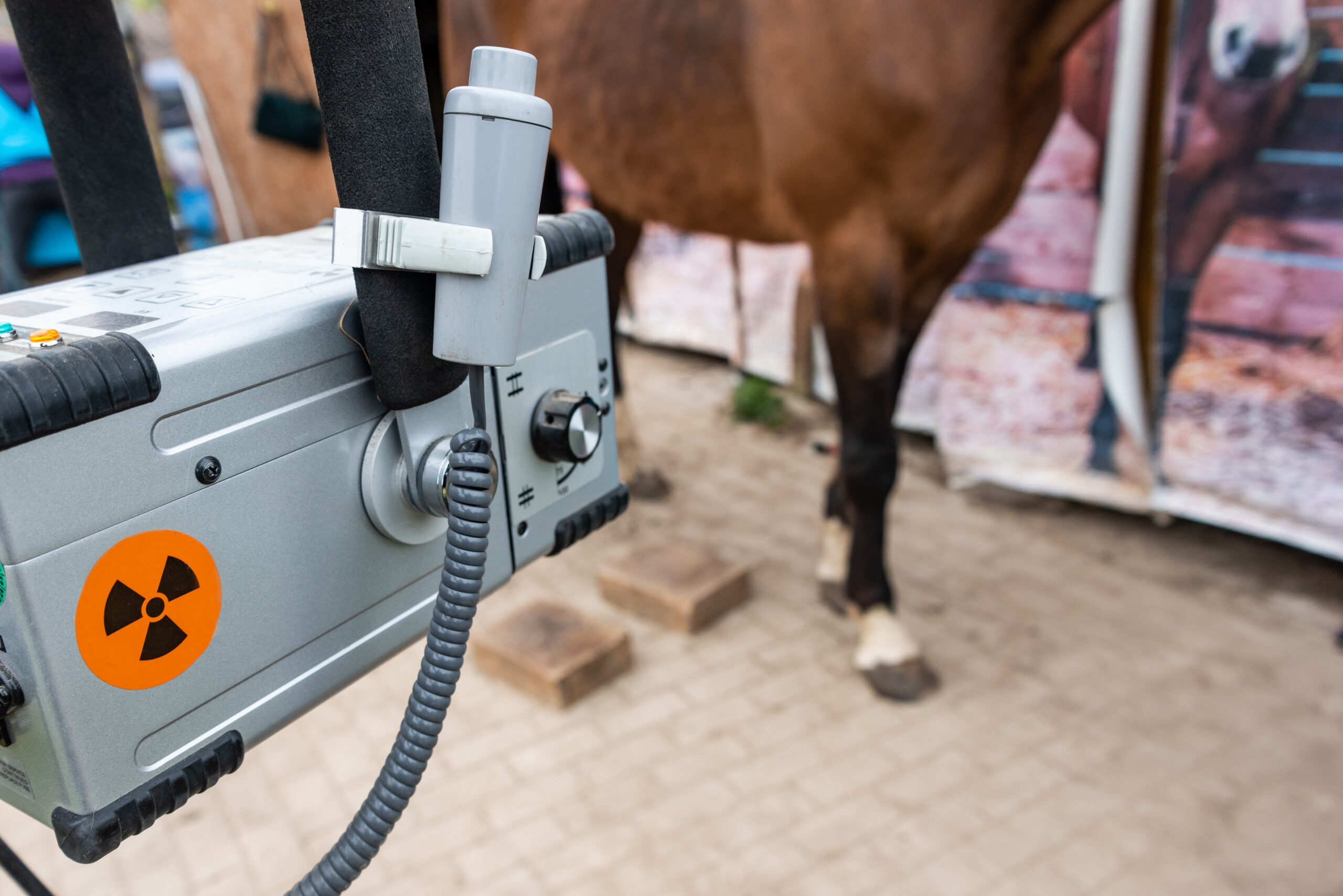
To obtain a radiograph, an X-ray generator emits and focuses a beam of X-rays toward the part of the horse’s body that is to be imaged. A detector on the opposite side of the area to be imaged detects the X-rays that pass through.
Dense structures like bone absorb more radiation. They stop the X-rays and appear white on the digital screen, while softer tissues allow more rays to pass through, showing up in various shades of grey. Air offers very little resistance to X-rays, so the area around the body (and areas of gas within the body) appear darker on the image.
Nowadays, almost all detectors are digital. Portable generators and detectors mean veterinarians can take and display most radiographs instantly at the yard, allowing them to check the quality of the images and see major findings right away. However, to scrutinise the finer details of radiographs, vets will often review them in a dark environment away from direct sunlight and without the hustle and bustle of a yard. Whilst portable generators are powerful enough to image most parts of the body, some areas require the higher power of a hospital-based generator to provide images of acceptable quality.
The Challenges of Taking Radiographs in Horses
Capturing a high-quality X-ray image of an animal as large as a horse presents several challenges that do not exist with smaller household pets or people. For the safety of everyone involved and to minimise blur due to movement, veterinarians usually give the horse mild sedation. This is safe for the vast majority of older horses.
Once the horse is calm, the veterinarian must work around the inherent limits of the technology itself. An X-ray is fundamentally a two-dimensional picture of a three-dimensional structure. Because bones overlap and shadow one another on a flat image, a single snapshot rarely tells the whole story. To overcome this, veterinarians must take multiple shots from different angles, known as orthogonal views, usually at 90-degree offsets from one another.
Furthermore, the size of each image is limited to a relatively small area by the size of the detector plate and focus of the X-ray beam. Whilst it is possible to capture the whole of a small animal such as a cat on a single detector plate, this isn’t possible with a horse. The veterinarian must carefully position different plates to capture the areas of interest. In some cases (such as during some prepurchase examinations) 30-40 different images of a horse may be obtained.
The size of the horse presents further challenges. Even with powerful generators, there are physical limits to what can be imaged. The massive size of a horse’s abdomen or pelvis is often too thick for the X-ray beam from many generators to penetrate, causing the beam to become scattered and limiting image quality. In some settings these areas can be imaged with more powerful hospital-based generators, but there remain significant limitations to the areas that can be imaged and the image quality that is feasible.
Inside the Skeleton: What X-Rays Can and Cannot Show
To make the best decisions for your senior horse, it is helpful to understand the strengths and limitations of X-rays. Radiographs are relatively quick to obtain and often provide good-quality images of hard mineral-rich structures such as bones, joint margins, dental roots, and metal or stone foreign bodies. They allow veterinarians to inspect bone density, spot fractures, and identify bone remodeling. X-rays are particularly useful in areas with high radiographic contrast. In these areas, there’s a significant difference in how X-rays are absorbed by one structure compared with surrounding areas such as the edges of bones at joint margins.
However, radiography has a major limitation when it comes to differentiating soft tissues such as tendons and ligaments. The radiographic appearance of many soft tissues does not materially change when they are damaged; swelling can sometimes be appreciated depending on the setting and the radiographic appearance of the surrounding area, but in general terms soft-tissue resolution is poor. Exceptions to this rule include changes that can occur at the sites where ligaments attach to bones. In areas of limited radiographic contrast, veterinarians will sometimes use special (high-contrast) liquids to highlight specific structures, such as joints or tendon sheaths.
It can also be difficult to detect early or mild changes on X-rays. This is a common problem following an impact such as a kick in which a fracture may be suspected. Cracks in the bone surface that don’t lead to displacement of a fragment of bone might be invisible on X-rays taken immediately after the impact but will be seen most easily on X-rays taken about 10 days later.
Common Senior Horse Conditions Where X-rays Are Useful
Despite these limitations, X-rays remain an important diagnostic tool for common conditions in senior horses. Osteoarthritis is very common in older horses, and bony changes can reflect years of wear and tear in the joints. Vets might look for narrowed joint spaces, bone spurs, or other forms of bone remodeling to identify arthritic joints. However, a certain amount of wear and tear is entirely normal in an older horse, and veterinarians must interpret their findings with care.
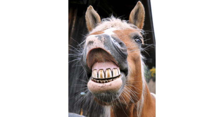
Dental disease is also common in older horses. By taking skull radiographs, veterinarians can identify problems with tooth roots and conditions such as sinusitis. The paranasal sinuses of the skull are usually filled with air through which X-rays pass easily; fluid such as pus in the sinuses can be clearly highlighted using X-rays.
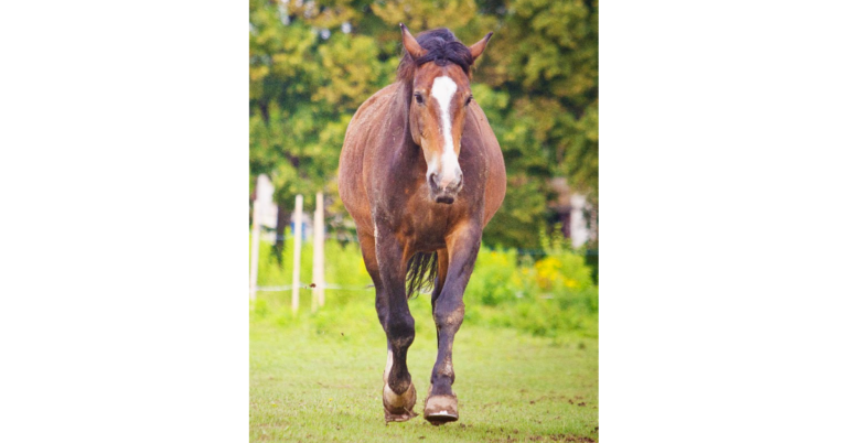
Laminitis is another common condition of older horses for which X-rays can be invaluable to reveal displacement of the pedal bone relative to the hoof capsule and remodeling of the pedal bone as a result of long-term disease.
When Your Veterinarian Might Recommend X-Rays
Radiographs can be useful for diagnosing many conditions. Common clinical signs in older horses that may lead to a recommendation for X-rays include:
- Persistent or worsening lameness.
- A swollen or painful joint.
- Suspected fracture after a fall or kick.
- Chronic or recurrent laminitis.
- Quidding, foul-smelling nasal discharge, or other signs of dental disease.
- Monitoring progression of arthritis or laminitis.
Looking Beyond X-Rays
X-rays are useful tools for imaging the horse, but other diagnostic imaging options will be more useful in some settings. For example, ultrasound can be more useful for investigating soft-tissue problems. Veterinarians often use ultrasound to evaluate tendons and ligaments or to investigate the intestines during episodes of colic.
For complex areas like the head, teeth, or lower limbs, particularly within the hoof capsule, advanced technologies like computed tomography (CT) or magnetic resonance imaging (MRI) are often needed. These modalities provide three-dimensional, cross-sectional views that capture both bone and soft tissue in great detail and can reveal a number of conditions that X-rays cannot.
Looking Beyond the Picture
X-rays can reveal many findings that may look dramatic but won’t cause the horse any problems. An important rule of veterinary medicine is to treat the horse, not the X-ray. Some horses remain completely sound and comfortable despite advanced bony changes on X-rays, while others are affected by pain in areas that appear pristine on X-ray. Interpreting X-ray changes is an important skill and images should be considered in the context of thorough examination of the horse and understanding of any previous veterinary findings.
Related Reading
- What Is Osteoarthritis in Horses?
- My Senior Horse Podcast: Senior Hoof Care
- Radiographic Abnormalities of Equine Neck Vertebrae
Sign up for My Senior Horse’s FREE newsletter to get the latest information about equids 15 years and older delivered straight to your inbox!